Shoulder Xray Interpretation Radiology Geeky Medics
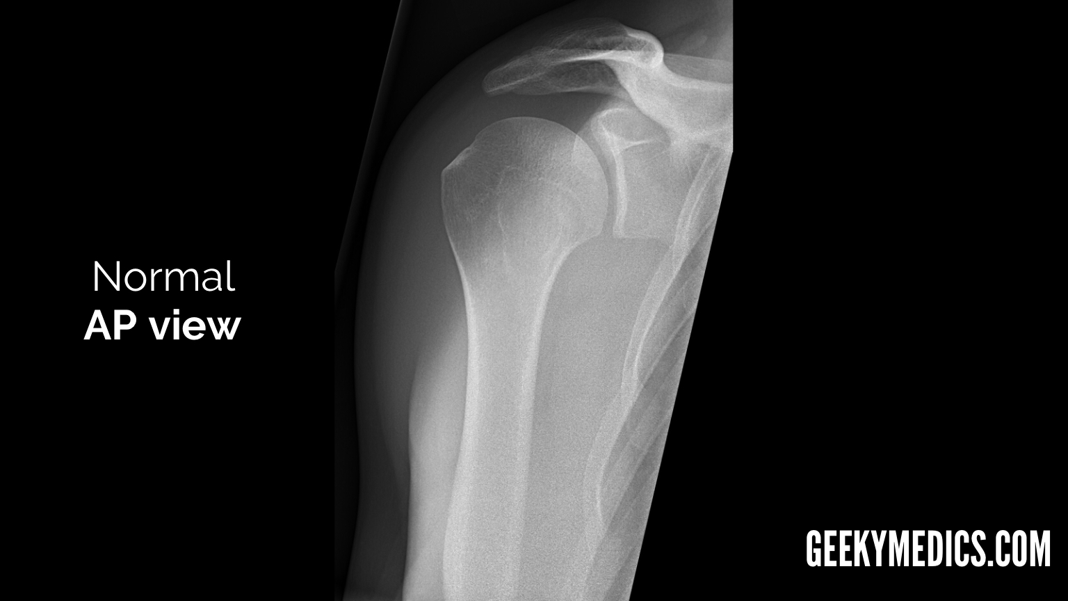
Typical shoulder X-ray views include: Antero-posterior (AP) view Lateral/scapula Y view (named due to the "Y" shape of the scapula in this view) An axial view can also be used as an alternative to the scapula Y view if the patient is unable to tolerate the positioning required to obtain this view. Figure 1. A normal AP view 1 Figure 1.1.
Shoulder X Ray Views slidesharedocs
The shoulder series is fundamentally composed of two orthogonal views of the glenohumeral joint including the entire scapula. The extension of the shoulder series depends on the radiography department protocols and the clinical indications for imaging. Indications Shoulder radiographs are performed for a variety of indications including:

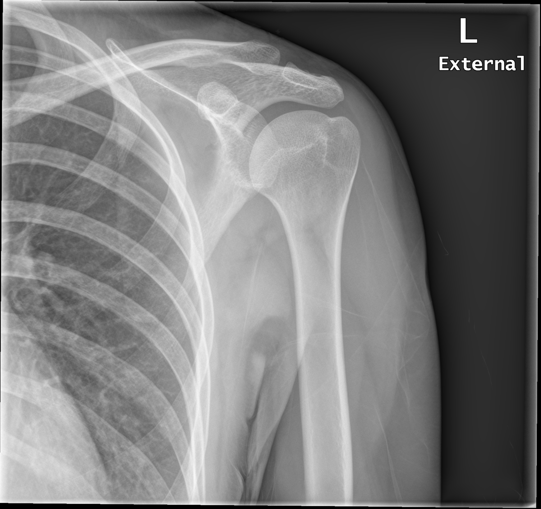
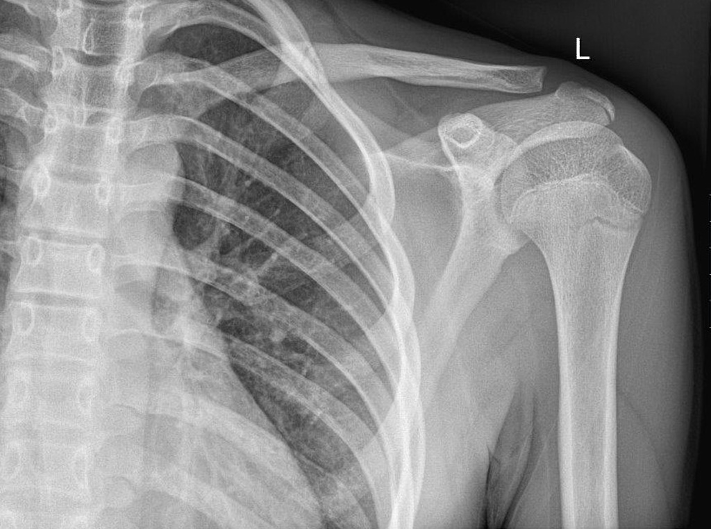
Right shoulder internal rotation and external rotation radiographs
Normal shoulder x-rays, including internal and external rotation views. 3 articles feature images from this case 43 public playlists include this case Related Radiopaedia articles Normal upper limb imaging examples Shoulder

in Shoulder Radiology Musculoskeletal Key
An X-ray of the shoulder is a frequently conducted examination and is mainly used for diagnosing a fracture. Some of the key topics are proximal humeral fracture, shoulder dislocation, Bankart lesion and osteoarthritis. KEY TOPICS/TERMS: Proximal humeral fracture Shoulder dislocation Hill-Sachs lesion Bankart lesion Osteoarthritis
Dislocation / Instability — Dr. R. Edward Glenn, Jr.
Normal shoulder joint. The 'shoulder' joint is more accurately termed the glenohumeral joint. In the context of trauma there are 2 standard views used to assess this joint. These are the - Anterior-Posterior (AP) view, and the lateral or 'Y-view'. If the patient can tolerate holding the arm in abduction, an 'axial' view is an alternative to the.
Case Study Joint Arthritis Management in 60 yr. Old Male
RR shoulder by Ahmed Magbari; MSK by Dimitrius N. J. Stamoulis; Shoulder by Dr. Rajesh Gothi; Shoulder X-Ray by Ahmed Mohamed Mohamed Eid Ali; Normal Radiographic Anatomy by Ashley Hook; Normal Anatomy by Matthew McGee; Anatomy by Muhammad Bin Zulfiqar; Anterior shoulder dislocation by Alexey; Anatomy by Merazul Alam; EXAMEN by Jose Ignacio Aragon

Normal Shoulder X Ray Normal Shoulder Girdle Orthopedic Chest Stock
Normal radiographic measurements of the shoulder are important in the evaluation of the osseous relationships in plain radiography. Normal measurements do not rule out pathology and must be considered in the context of other findings and the clinical presentation. acromioclavicular (AC) joint space: 1-7 mm 4 (narrower in the elderly)

Shoulder Xray Anatomy
MRI is best for evaluating soft tissue structures and evaluating bone contusions or trabelcular microfractures. the stronger the magnet, the higher the intrinsic signal-to-noise ratio (e.g. a 3 Tesla MRI machine has 9x the proton energy of a 1.5 Tesla MRI machine) T1-weighted sequence. uses a short repetition time (TR) and short echo time (TE)

Normal Shoulder X Ray Normal Shoulder Girdle Orthopedic Chest Stock
While achieving anteroposterior shoulder X-ray in neutral position, the patient is erect or in supine position. Central X-ray should be directed to 2.5 cm inferior to the coracoid process.. The important anatomical structures of the normal shoulder joint are shown in axial, coronal, and sagittal CT images below (Figs. 3.8a-s, 3.9a-j, and.
NORMAL SHOULDER 7
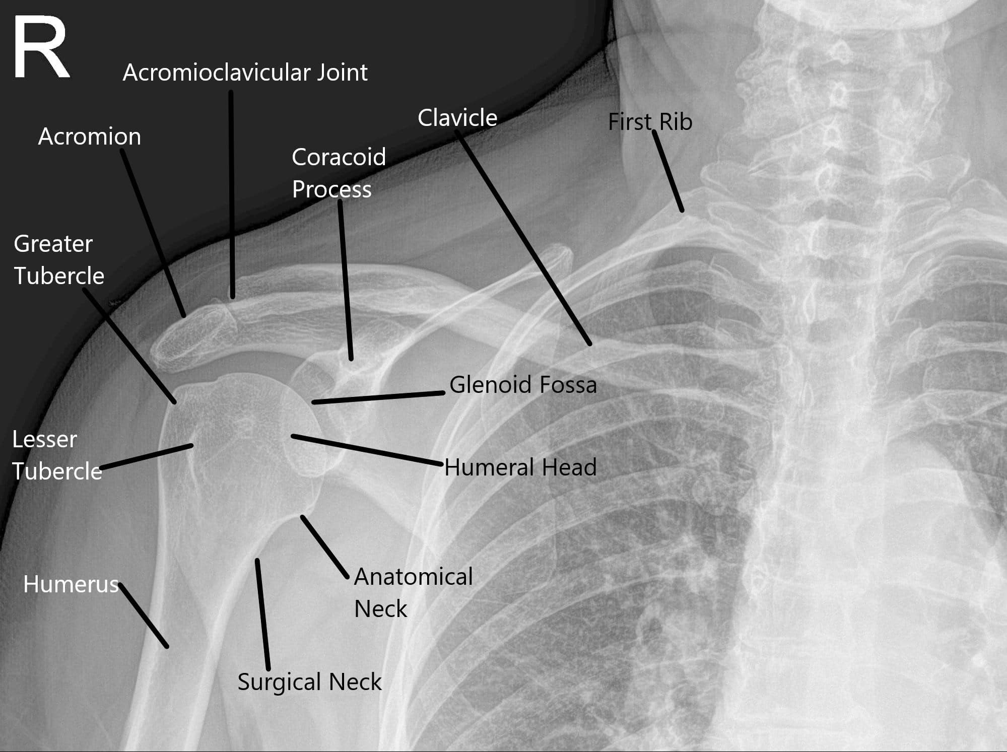
A normal shoulder X-ray will show the bones that make up this ball-and-socket joint: Humerus (upper arm bone). Scapula (shoulder blade), which connects to the humerus. Acromion (a piece of bone that projects off the scapula). Clavicle (collarbone), which connects to the acromion.

Image
A shoulder X-ray is a non-invasive imaging technique that utilizes a small dose of ionizing radiation to create detailed images of the shoulder joint. This diagnostic tool is crucial for diagnosing a wide range of shoulder conditions, such as fractures, dislocations, arthritis, and more.
Normal Shoulder X Ray Xschouder Startpuntradiologie.nl Arm is
A normal humeral head looks like a walking stick on the AP view. The most common fracture of the humerus is a metaphyseal fracture. Metaphyseal fractures occur in ages 5-12 and Salter-Harris fractures outside of this range. Don't forget the scapula, seen best on the Y view.

Normal shoulder, Xray Stock Image F003/9192 Science Photo Library
A video tutorial in interpreting radiographs of the shoulder joint and surrounding areas. This is the second video in a series of five by TeachMeAnatomy -- h.

AP of the glenohumeral joint Medical anatomy, Radiology schools
Normal shoulder X-ray A normal shoulder x ray will demonstrate the bones of the shoulder to have expected normal appearance without breaks, bone lesions, or abnormal bone structure. The head of the humerus or upper arm will be positioned within the socket of the shoulder.

Image
Normal and Variant Anatomy by James Clark Normal Radiographs by Osamah A. A. Alwalid; Nicole's shoulder and pelvis II playlist by Denise Foulkes; MSK by Johann Jende; MSK by Naveed Ahmad; Normal radiographs by Leonardo Lustosa Normal, Anomalies & Dysplasias by Varsha Kumar; Membre supérieur by Laurence; rad club april 27 by Anser Abbas

Shoulder & Knee Doc
An approach to the traumatic adult shoulder x-ray The American College of Radiology recommends at least 3 views for acute traumatic shoulder pain [5]: AP in internal rotation for visualization of the lesser tuberosity AP in external rotation for visualization of the greater tuberosity Scapula Y or axillary view in place of true lateral.